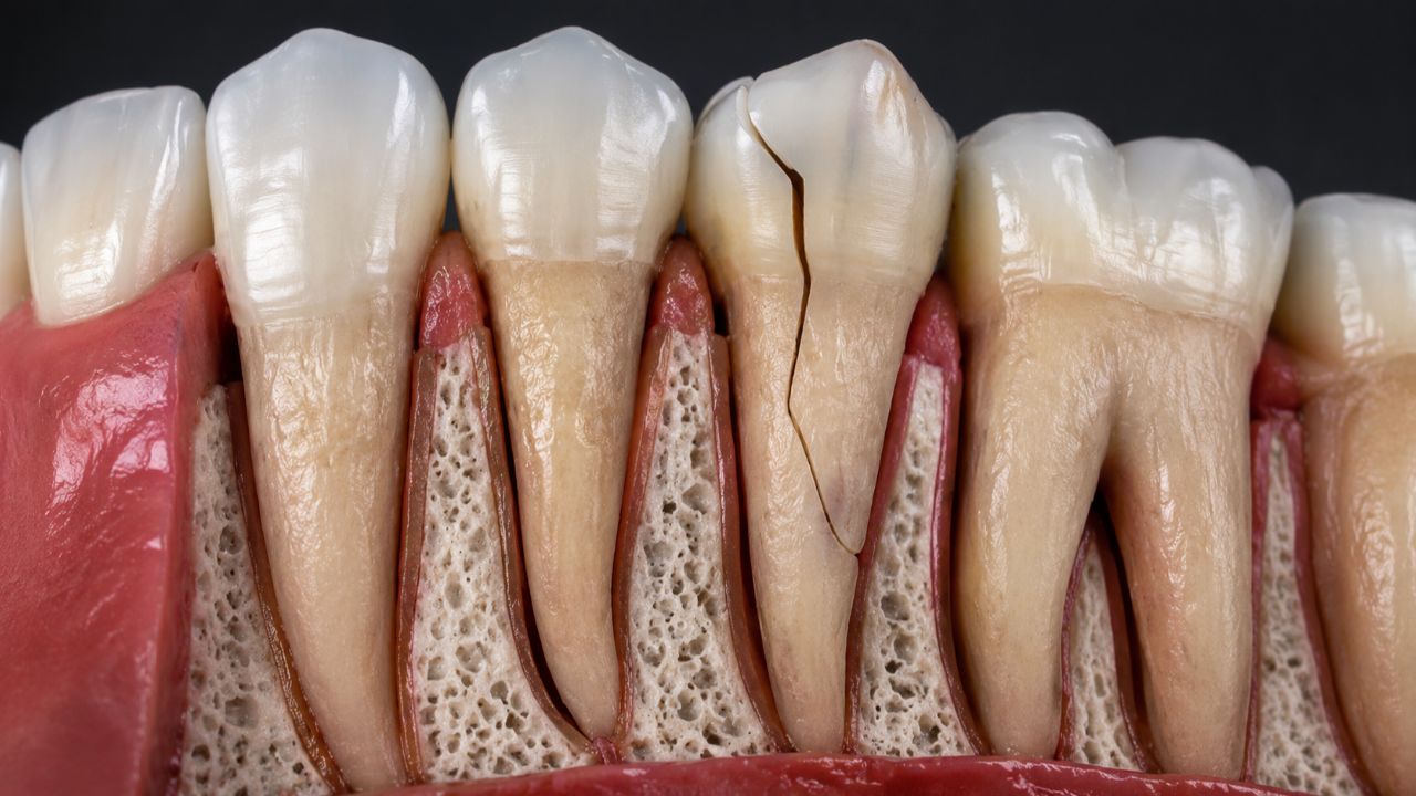
A vertical root fracture is one of the most difficult conditions to diagnose in Endodontics. It may cause only minor symptoms, mimic persistent infection and, in its early stages, may not be directly visible on radiographs or even on cone beam computed tomography (CBCT).
This matters because the diagnosis has major consequences: in many cases, a vertical root fracture seriously compromises the prognosis of the tooth.
What is a vertical root fracture?
It is a crack or fracture that develops longitudinally along the root.
It may be complete or incomplete and extend in different directions. As bacteria gain access through the fracture, localised inflammation of the periodontal tissues and bone loss alongside the root may develop.
It is more frequently discussed in previously root canal-treated teeth, although it can occur in other situations.
Why can it look like a failure of root canal treatment?
A tooth with a vertical root fracture may present with:
- mild pain or discomfort when chewing;
- a recurrent sinus tract;
- localised bone loss;
- gingival inflammation in a restricted area;
- a radiographic lesion alongside the root;
- symptoms after apparently adequate root canal treatment.
These findings can also occur with an untreated canal, a perforation, persistent infection or a periodontal problem.
For this reason, when there is pain in a previously treated tooth, fracture is only one of several possible diagnoses that need to be excluded.
Is there a typical sign?
A very deep, narrow periodontal probing defect at one isolated point can raise strong suspicion, particularly when the rest of the periodontium is healthy.
Certain patterns of bone loss can also be suggestive, such as narrow defects extending along the side of the root.
But none of these findings is specific to a fracture. Diagnosis results from combining the available information.
Does CBCT always show the fracture?
No.
This is one of the most common misconceptions.
CBCT is extremely useful in Endodontics, but a fracture line may be thinner than the resolution of the scan. Root-filling materials, metal posts and other restorations can also create artefacts that make interpretation more difficult.
Recent scientific reviews continue to show that the diagnostic performance of CBCT for vertical root fractures varies substantially and is less reliable in previously treated teeth.
Therefore, not seeing a fracture on CBCT does not prove that it is absent.
So what is CBCT useful for in these cases?
Often, the most useful information is not direct visualisation of the fracture line, but its consequences.
Three-dimensional imaging can show patterns of bone loss, the location of the lesion and its relationship with adjacent roots, while also helping exclude other causes such as untreated canals, perforations or anatomical variations.
At the Portuguese Institute of Endodontics, we use small-field-of-view CBCT when there is a specific clinical indication. Technology should answer a question, not replace diagnosis.
What about the microscope?
The operating microscope may allow cracks to be seen in the crown, on the floor of the pulp chamber or on accessible root surfaces.
However, part of the root is hidden by bone and periodontal tissues. The suspicious area cannot always be inspected directly without surgery or extraction.
In some cases, definitive confirmation is only obtained through surgical exploration when that information could change the treatment decision.
Why do these fractures occur?
There is usually no single cause.
Loss of tooth structure, large restorations, root anatomy, posts, occlusal forces and previous procedures can all influence risk.
It is incorrect to say that root canal treatment by itself “makes the root brittle”. As explained in “Does Root Canal Treatment Weaken a Tooth? Myth or Fact?”, the amount of remaining tooth structure is a central factor.
Can a vertical root fracture be treated?
It depends on the tooth and the location of the fracture.
In a single-rooted tooth, a true vertical root fracture generally has a poor prognosis and frequently leads to extraction.
In some multi-rooted teeth, if the fracture is confined to one root and the remainder of the tooth is suitable, more conservative options may exist in carefully selected cases.
The decision must also take restorability, periodontal support and the strategic value of the tooth into account.
Why is it risky to diagnose too early — or too late?
If a fracture is assumed without sufficient evidence, a potentially treatable tooth may be extracted unnecessarily.
If, on the other hand, an obvious fracture is ignored and repeated Endodontic procedures are performed without addressing the cause, the patient undergoes treatment with no predictable benefit.
The balance lies in reaching a sufficiently robust diagnosis before an irreversible decision is made.
At the Portuguese Institute of Endodontics, teeth with suspected fractures are assessed using detailed clinical examination, probing, magnification and imaging when indicated. The objective is not to “save the tooth at any cost”, but to determine whether a realistic and predictable alternative to extraction exists.
If you have received a diagnosis of vertical root fracture and have questions, it may be reasonable to seek a second opinion before extracting the tooth.