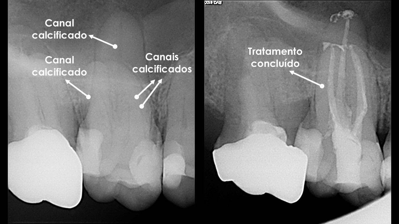
When a root canal is heavily calcified, the space normally occupied by the pulp can become extremely narrow or appear to disappear on radiographs.
This can make root canal treatment significantly more demanding, but a calcified canal does not automatically mean that the tooth is lost or impossible to treat.
What does it mean to have a calcified root canal?
Throughout life, the pulp may deposit dentine inside the tooth. This process can be accelerated by ageing, trauma, decay, extensive restorations or other stimuli.
As a result, the pulp chamber and root canals progressively become narrower.
In advanced situations, the canal may become almost invisible on a conventional radiograph, although a microscopic space often remains within the root.
Does a calcified canal always require treatment?
No.
Calcification alone is not an indication for root canal treatment.
A calcified tooth may remain asymptomatic and healthy for many years. Treatment is considered when there is pulpal or periapical disease, symptoms, infection or another specific clinical indication.
We should treat disease, not simply a radiographic appearance.
Why are these cases more difficult?
In a tooth with normal anatomy, canal entrances are identified using familiar anatomical landmarks.
When calcification is present, these landmarks may be altered or covered by dentine. Searching for the canal without a precise strategy increases the risk of unnecessarily removing tooth structure, creating a perforation or deviating from the original canal path.
This is why these cases are considered more difficult.
How does the operating microscope help?
The operating microscope provides magnification and intense illumination of the treatment field.
It can help distinguish subtle colour and texture differences in dentine, identify traces of the original anatomy and allow much more controlled work.
The advantage is not simply “seeing larger”. It is being able to remove only the dentine that is necessary while searching for a space that may be extremely small.
What about CBCT?
In selected cases, small-field-of-view cone beam computed tomography (CBCT) can help assess the likely position and direction of the canal in three dimensions.
It is particularly useful when calcification is extensive and there is a risk of losing orientation during access.
At the Portuguese Institute of Endodontics, we have high-resolution, small-FOV CBCT for situations in which this information may alter the treatment strategy.
CBCT is not necessary for every calcified tooth. It should be requested when it provides relevant additional information.
Which techniques can be used to locate the canal?
In addition to magnification, very fine ultrasonic instruments can be used to remove dentine in a controlled manner.
The canal is explored progressively, often using delicate hand instruments, with the aim of recovering the original pathway without weakening the root.
In cases of extreme calcification, guided Endodontic techniques using three-dimensional planning can also be considered. They are not necessary in every case and have specific indications.
Can the canal be completely closed?
Radiographically, it may appear so, but a two-dimensional image does not reveal every microscopic detail.
Even when the canal appears obliterated, a very fine space may remain. In some cases, however, negotiating the canal to its full length may not be possible or may involve excessive risk.
The decision must balance benefit and safety. Forcing access at any cost is not good Endodontic practice.
What happens if it is not possible to reach the end of the root?
It depends on the diagnosis and the location of disease.
If persistent infection is present in an area that cannot be reached conventionally, another strategy may need to be considered, including monitoring or Endodontic microsurgery in selected cases.
In previously treated teeth, it may also be necessary to assess whether root canal retreatment or another approach is appropriate.
A tooth that calcifies after trauma requires particular attention
Trauma can trigger progressive pulp calcification, sometimes accompanied by a change in tooth colour.
Many of these teeth remain vital and do not require treatment. Others may later develop necrosis and periapical disease.
Follow-up is therefore important. The article on dental trauma explains why some problems only become apparent months or years later.
Experience and planning make a difference
A calcified canal is a good example of how Endodontic difficulty depends more on anatomy than on the apparent size of the problem.
At the Portuguese Institute of Endodontics, these cases are assessed using microscopy, radiography and CBCT when indicated, with the aim of achieving a conservative and predictable approach.
If you have been told that a canal is “closed” or impossible to treat, an assessment by an endodontist can help determine what options genuinely remain.