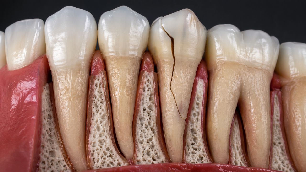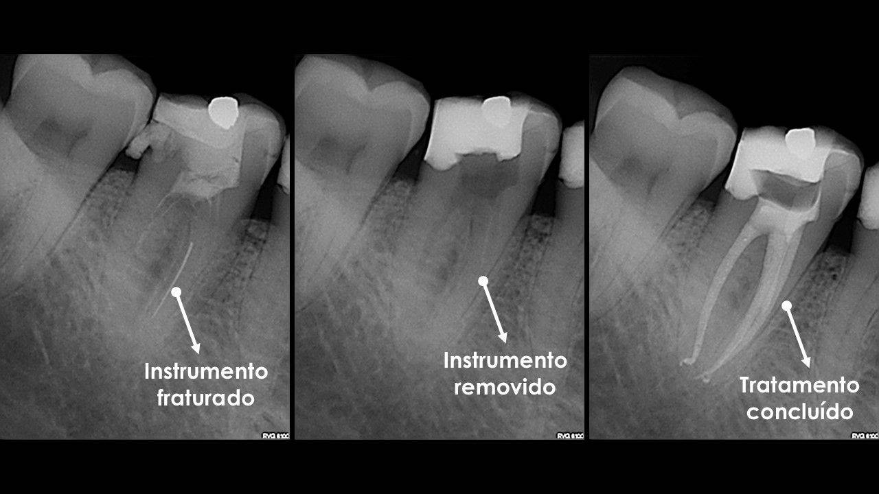
During root canal treatment, extremely fine instruments may be used to clean and shape the root canals. In some situations, one of these instruments may fracture and remain lodged inside the canal.
For the patient, this is understandably worrying news. However, it is important to understand that a separated instrument does not automatically mean that the tooth is lost or that the treatment has necessarily failed.
Today, there are strategies and devices specifically developed to manage these situations. In many cases, it is possible to bypass the fragment or remove it, restoring access to the remaining root canal system and allowing disinfection and treatment of the tooth to be completed.
The impact depends mainly on the initial diagnosis, the location of the fragment, the root anatomy and the degree of disinfection achieved before the instrument separated.
Why can an instrument break?
Root canals can be narrow, curved and anatomically complex. During instrumentation, the instruments are subjected to torsional forces and repeated flexion.
Even with appropriate protocols, inspection and regular replacement, there is an inherent risk of instrument separation.
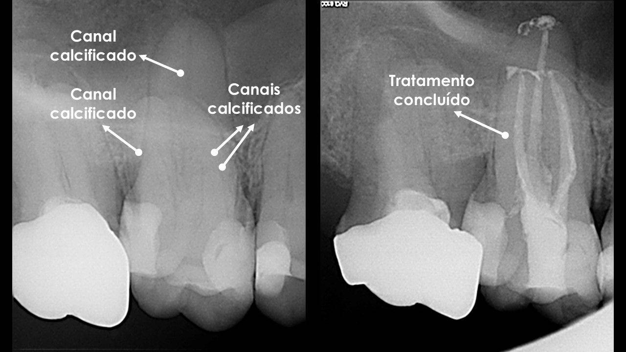
Severe curvatures, calcified canals, excessive reuse, unexpected anatomy and instrument fatigue are some of the factors that may influence this risk.
Is the fragment an infection?
No.
The instrument itself is an inert material. The problem is not the metal, but the possibility that the fragment may block access to a portion of the canal that still contains bacteria or contaminated tissue.
For this reason, its clinical significance can be very different in a vital tooth treated because of pulpal inflammation compared with a tooth with pulp necrosis and an extensive periapical lesion.
The priority is to regain access to the canal: bypass or removal
When a separated instrument is present, one of the first questions is whether it can be bypassed or safely removed.
If the fragment is in an accessible area and there is sufficient tooth structure around it, removal may allow the original canal pathway to be fully recovered.
In other cases, it may be possible to perform a bypass. This means carefully passing very fine instruments alongside the fragment and regaining access to the apical region. Once a bypass has been achieved, it becomes possible to irrigate, disinfect and shape the canal beyond the separated instrument. In some cases, the fragment itself becomes mobile and can subsequently be removed.
The strategy depends on the position of the instrument, the length of the fragment, the curvature of the root, the thickness of the remaining dentine and the experience of the clinician.
There are situations in which an aggressive attempt at removal would create more risk than benefit. However, that decision should only be made after carefully assessing whether access can be regained through bypass or removal.
How can a separated instrument be removed?
Removing a fragment can be one of the most technically demanding procedures in Endodontics.
It is not simply a matter of “pulling” the instrument out. It is often necessary to create space around the coronal end of the fragment, progressively free it and control its movement without removing excessive dentine.
This may require equipment and instruments specifically designed for fragment removal, including:
- specific ultrasonic tips, which are extremely fine and used under magnification to create space and transmit controlled vibration to the fragment;
- microtube systems, which can engage or capture the end of the instrument and apply controlled traction;
- wire loop systems, used in selected situations to secure the fragment and facilitate its removal.
The choice of technique depends on the anatomy and the position of the instrument. Sometimes a combination of several techniques is required during the same procedure.
These devices can resolve situations that would be extremely difficult using conventional instrumentation, but they must be used with great control. Excessive removal of root structure may weaken the tooth, increase the risk of perforation or create a complication more difficult than the original problem.
For this reason, these systems should be used by an endodontist experienced in complex procedures for the removal of separated instruments.
What does it mean to bypass the fragment?
Bypassing means successfully passing very fine instruments alongside the fragment and regaining the original canal pathway beyond it.
This technique can be particularly useful when the instrument cannot be removed immediately or when direct removal would require excessive removal of tooth structure.
A bypass makes it possible to reach the apical part of the canal again, irrigate and disinfect beyond the fragment, and complete preparation of the root canal system.
In some cases, once the bypass has been established, the fragment becomes more mobile and can be removed at a later stage. Bypass and removal are therefore not necessarily opposing strategies. They may form part of the same treatment sequence.
Not every fragment can be bypassed, but when there is a reasonable technical possibility, this approach can be decisive in recovering treatment of the canal without compromising the root.
How can the operating microscope help?
The operating microscope is particularly important in these procedures.
Magnification and illumination make it possible to identify the end of the fragment, work in extremely small areas and use ultrasonic tips, microtubes and other devices with much greater control.
It also allows the clinician to continuously assess how much tooth structure is being removed and adjust the strategy before unnecessarily compromising the root.
In a procedure where differences of fractions of a millimetre can alter the prognosis, the ability to see and work precisely is essential.
What about cone beam computed tomography?
Cone beam computed tomography (CBCT) can be particularly useful when planning these cases because it allows the situation to be studied in three dimensions.
A conventional radiograph provides a two-dimensional projection. The fragment may appear to be in a certain position, but the radiograph cannot directly show whether the root curves in the buccolingual direction or whether there are other three-dimensional anatomical features.
With CBCT, it is possible to assess exactly where the fragment is located within the root, its relationship with canal curvatures, the amount of dentine surrounding it and the likely pathway beyond it.
This is especially important when the fragment is located within a curvature that is not visible on a two-dimensional radiograph. This information can completely change the decision regarding the best direction of access, the feasibility of bypass and the risks associated with an attempt at removal.
CBCT may also help assess root anatomy, dentine thickness and the relationship between the fragment and any periapical lesion.
This three-dimensional information allows the intervention to be planned before treatment begins and supports a more conservative strategy.
Does a separated instrument always reduce the chance of success?
Not necessarily.
The prognosis depends much more on the presence and control of infection than on the physical presence of the fragment itself.
When the fragment can be bypassed or removed and access to the root canal system is regained, disinfection and treatment of the previously blocked region can continue.
This is precisely why early identification of the fragment and a specialist approach can be so important, particularly when pulp necrosis or periapical disease is present.
What if the tooth was treated previously and contains an old fragment?
If there is pain in a previously treated tooth, persistence of a lesion or signs of infection, it is important to assess whether the fragment is preventing access to a contaminated area and whether root canal retreatment may improve the prognosis.
In these cases, removing or bypassing the instrument may be an essential step in regaining access to a portion of the canal that was not adequately treated.
When a tooth is completely asymptomatic, shows normal healing and there are no signs of disease, there may be situations in which intervention is not indicated simply because a fragment is visible on a radiograph. However, this is an individual clinical decision and should not be assumed without proper assessment.
What if the fragment cannot be removed or bypassed?
Despite all the available resources, some fragments are positioned in such a way that their relationship with the anatomy makes bypass or removal excessively risky.
In these situations, the decision depends on the diagnosis and on whether disease is present beyond the fragment. It may be appropriate to complete treatment as far as possible and monitor the tooth or, if persistent disease is present at the root end and cannot be reached through conventional treatment, to consider endodontic microsurgery.
Extraction is reserved for situations in which the tooth cannot be predictably treated or restored, not simply because a separated instrument is present.
At the Portuguese Institute of Endodontics, these cases are part of our clinical practice
The Portuguese Institute of Endodontics is a clinic with a particular focus on highly and extremely complex endodontic cases.
We have the different resources used in these procedures, including operating microscopy, dedicated ultrasonic tips, microtube systems and wire loop systems, allowing us to select the most appropriate strategy for each situation.
Prof. Rui Pereira da Costa has dedicated his clinical practice exclusively to Endodontics for more than 20 years and has accumulated extensive experience in managing complex cases, including numerous separated-instrument cases resolved through bypass, removal or a combination of different techniques.
This experience is particularly important because two fragments that appear similar on a radiograph may require completely different approaches once the three-dimensional anatomy, canal curvature and available tooth structure are taken into account.
The goal is to recover treatment without creating a bigger problem
Removing an instrument should not mean removing dentine indiscriminately. The goal is to regain access to the canal as conservatively as possible, whether through bypass, direct removal or a combination of both strategies.
When technically possible, this allows the previously blocked region to be treated and improves the ability to control infection without sacrificing the tooth.
At the Portuguese Institute of Endodontics, we assess these cases using magnification, CBCT when indicated and dedicated instrumentation in order to plan the strategy as precisely as possible.
If you have been told that a tooth contains a broken instrument and needs to be extracted, it may be worth seeking a specialist second opinion before making an irreversible decision.